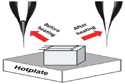
Researchers at Kanazawa University report in Analytical Chemistry an efficient method for filling a batch of nanopipettes with a pore opening below 10 nanometers. The method is based on the application of a temperature gradient to the nanopipette tips so that residual air bubbles are driven out.
Nanopipettes, in which a nanoscale channel is filled with a solution, are used in all kinds of nanotechnology applications, including scanning-probe microscopy. Bringing a solution into a nanopipette with a pore diameter below 10 nanometer is challenging, however, since capillary forces prevent the complete filling of a sub-10-nm nanopipette pore with a liquid. Now, Shinji Watanabe and colleagues from Kanazawa University have found a simple but efficient way for filling nanopipettes. The researchers show that the 'air bubble' that typically remains near the pipette's pore end can be removed by applying a temperature gradient along the pipette.
The scientists investigated their 'thermally-driven method' to a batch of 94 pipettes, aligned length-wise next to each other, all with a pore diameter of around 10 nm. The pipettes were put on a metal plate kept at a temperature of 80 °C, with their tips protruding from the plate, resulting in a temperature gradient.
Time-lapsed optical microscopy images of the filling process of the nanopipettes showed that after 1200 seconds, the tips are completely filled with solution, and that air bubbles are driven out of the pipettes.
In order to double-check that the pipettes were indeed bubble-free, Watanabe and colleagues performed so-called I–V measurements. Every pipette was filled with a solution of potassium chloride (KCl), which is conducting. Both pipette ends were then contacted with electrodes. If an electrical current runs between the ends—specifically, if the pipette has an electrical conductivity below a few GΩ— then filling with the solution is complete. The resesarchers observed electrical currents and therefore filling for the whole batch of pipettes.
The scientists also performed transmission electron microscopy (TEM) measurments of pipettes with pore diameters below 10 nm. Although the thermally-driven method leads to good electrical contacts, particle-like structures were observed inside the tips of the nanopipettes, demonstrating that (quoting the researchers) "TEM observation without inducing pipette deformation is important for accurately determining the characteristics of sub-10-nm nanopipettes."
Watanabe and colleagues concluded that their method "is very practical and easy to introduce in nanopipette frabrication" and that their study "will provide a significant contribution to various fields of nanoscience using nanopipettes".
Nanopipettes
Nanopipettes are usually made from quartz or glass, and have a pore opening in the nanometer range. Today, nanopipettes are used for molecular sensing, delivery of chemicals and scanning-probe microscopy. The latter is a technique for imaging a material's surface by scanning a probe over it; for the probe, a solution-filled nanopipette can be used.
The function of a nanopipette is usually to enable the transport, and their detection, of nanometer-sized objects (in solution) through the pipette pore.
Completely filling a nanopipette with a solution has been difficult: because of the capillary force, an 'air bubble' is nearly always present in the pipette's tip. Removing the air bubble has proven to be problematic for nanopipettes with a pore opening of 10 nanometer or less.
Now, Shinji Watanabe and colleagues from Kanazawa University have found a way to achieve complete filling of a batch of many nanopipettes with a pore opening of about 10 nm. The method, based on the application of a temperature gradient to the nanopipettes, is simple and efficient.

