Scientists from Utrecht University have applied advanced microscopy to visualize the activity of the widely used drug Taxol. Taxol is often used in cancer chemotherapy. A better understanding of the precise action of the drug offers starting points for less toxic therapies. The scientists, together with international colleagues, have published their findings in Nature Materials.
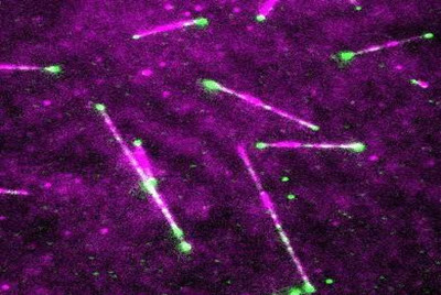
Taxol is a drug that is frequently used in chemotherapy. It acts on microtubules, cellular filaments that are involved in cell division and cell migration. "Microtubules resemble spaghetti tubes made up of small balls. By labeling microtubules and the drugs with fluorescent molecules, we can follow how microtubules are formed and what happens when the drug binds to them," says the last author Anna Akhmanova of Utrecht University. Using advanced microscopy, Utrecht researchers, in collaboration with British, Swiss, Spanish and Chinese colleagues, were able for the first time to observe the effects of Taxol on growing microtubules directly.
"The fluorescent label allowed us to see under the microscope that the drug binds to growing microtubule ends. As soon as one molecule binds, more start to bind, and a kind of hotspot is created. The 'tube' becomes more stable than usual at this location," Akhmanova explains. This is beneficial for the fight against cancer cells: The ability of cancer cells to divide and migrate is inhibited, which limits tumor growth and metastases.
Growth and shortening of microtubules, with fluorescent markers at the tips. Credit: Ankit Rai, Utrecht University.
To their surprise, the researchers also discovered that drugs that stabilize the cellular skeleton and destabilize it can have a synergistic effect. "We didn't expect this," says Akhmanova. "For the first time we see these drugs in action. That is very special. By looking at the combined effect of drugs that affect microtubules, we can work toward therapies that are effective, but less toxic."

